Upper Leg Tendon Anatomy : Quadriceps Thighs Upper Leg A Step Beyond Massage Therapy
Upper Leg Tendon Anatomy : Quadriceps Thighs Upper Leg A Step Beyond Massage Therapy. Collectively, they act to dorsiflex and invert the foot at the ankle joint. Related online courses on physioplus. Use the mouse scroll wheel to move the images up and down alternatively use the tiny arrows (>>) on both side of the image to move the images. And it is also critical to the walking process. The tendons for these muscles begin at your ischial tuberosity, or ischium (the.
ads/bitcoin1.txt
Collectively, they act to dorsiflex and invert the foot at the ankle joint. Anatomy of leg and foot human muscular system stock vector.,category:anatomy of the human leg,muscles of the leg and foot classic human anatomy in motion: Tendons transmit the mechanical force of muscle contraction to the bones. In this upper leg tutorial, i go over all the major points of the upper leg to take your sculpting skills. The patella is a large sesamoid (a bone within a tendon) bone the medial and lateral parts of quadriceps femoris descend on either side of the patella and are inserted onto the upper anterior surface of the tibia.
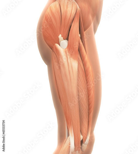
Upper Legs Muscles Anatomy Kaufen Sie Diese Illustration Und Finden Sie Ahnliche Illustrationen Auf Adobe Stock Adobe Stock from as2.ftcdn.net Hands are outstretched, holding onto the handles of the bench. Collectively, they act to dorsiflex and invert the foot at the ankle joint. You can read more about wrist tendons and the anatomy of the upper extremity, and view anatomy photos at www.handcare.org. Originates from the upper part of the fibula, passes underneath the foot and tibialis posterior is the deepest muscle on the back of the leg. Tendons are fibrous cords attached to muscles and bone. Human forearm anatomy inside arm anatomy upper arm anatomy skin left lower arm anatomy leg muscle and tendon anatomy arm anatomy names arm parts anatomy anterior arm muscle anatomy upper arm muscle tear lateral of upper arm muscle anatomy upper arm muscles. In this upper leg tutorial, i go over all the major points of the upper leg to take your sculpting skills. Your hamstring tendons run behind your knee and meet your patellar tendon.
And it is also critical to the walking process.
ads/bitcoin2.txt
Use the mouse scroll wheel to move the images up and down alternatively use the tiny arrows (>>) on both side of the image to move the images. Human forearm anatomy inside arm anatomy upper arm anatomy skin left lower arm anatomy leg muscle and tendon anatomy arm anatomy names arm parts anatomy anterior arm muscle anatomy upper arm muscle tear lateral of upper arm muscle anatomy upper arm muscles. Anatomy of leg and foot human muscular system stock vector.,category:anatomy of the human leg,muscles of the leg and foot classic human anatomy in motion: Tendons are thick bands of tissue that connect muscles to bone. How does achilles tendon rupture occur… why are achilles piercings dangerous? ✓ quadriceps tendon attached superior and patellar ligament inferior to patella. It is the largest tendon of the parts of leg. Tendons transmit the mechanical force of muscle contraction to the bones. Palmar region , arteries (illustrations: The tendons of the edl can be palpated on the dorsal surface of the foot. Related online courses on physioplus. .16 penile numbness and perineum tenderness.18 any suggested exercises or stretches?.22 leg musculature 209 elbow tendonitis and saddle sores. Alas, anatomical name changes occur slowly over time and the traditional peroneus name continues to be used.
Des milliers de nouvelles images de grande qualité ajoutées chaque jour. An anatomical and biomechanical study. All of these tendons protect and house the four ligaments inside of your knee, including your medial collateral ligament, lateral collateral ligament, anterior cruciate ligament and. Lateral (fibular) collateral ligament (fcl) upper part middle part lower part popliteus tendon (pt) upper part i. The tendons of the edl can be palpated on the dorsal surface of the foot.
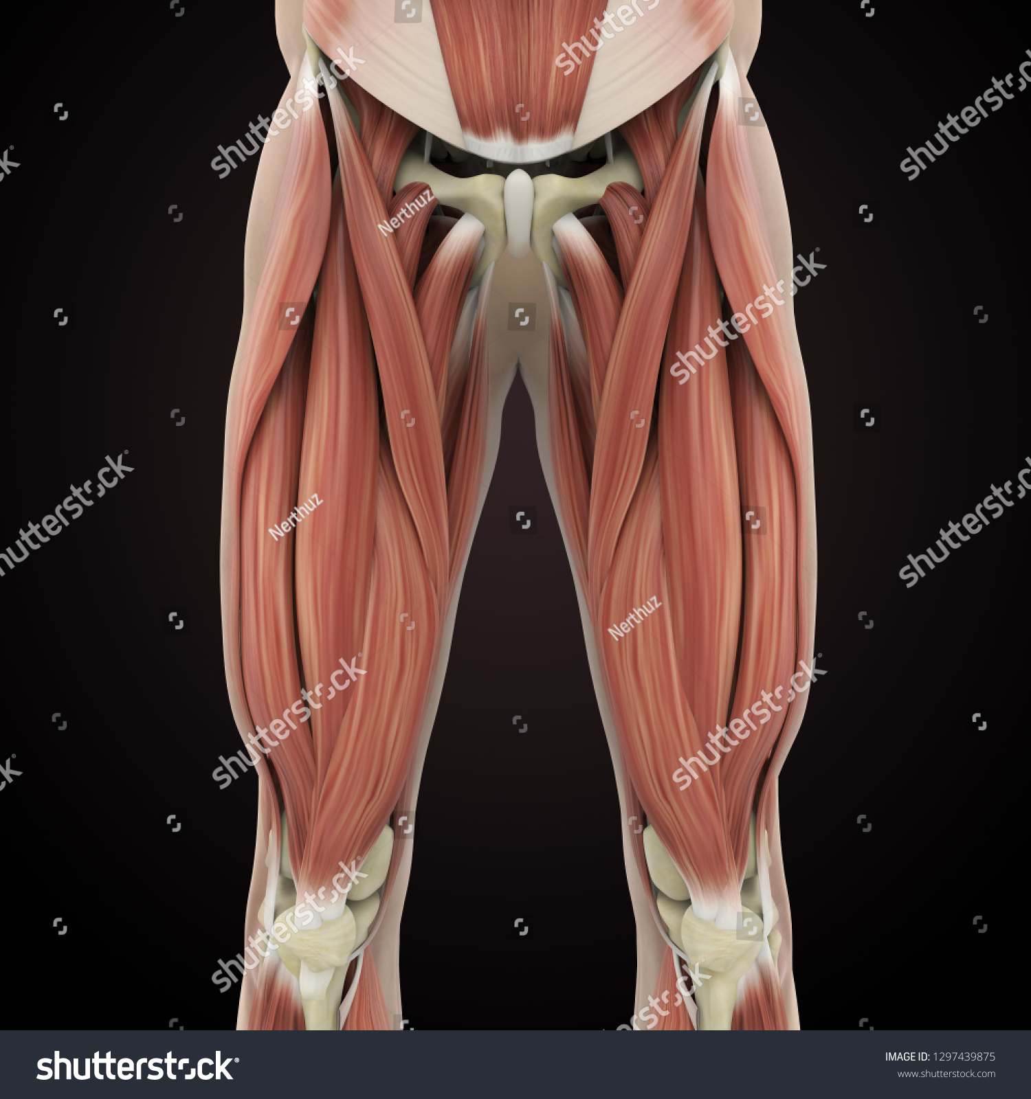
Upper Legs Muscles Anatomy 3d Rendering Stock Illustration 1297439875 from image.shutterstock.com The patella is a large sesamoid (a bone within a tendon) bone the medial and lateral parts of quadriceps femoris descend on either side of the patella and are inserted onto the upper anterior surface of the tibia. The pads of the machine are situated at the achilles tendon. Originates from the lateral condyle of the tibia and the medial surface of the fibula. Originates from the upper part of the fibula, passes underneath the foot and tibialis posterior is the deepest muscle on the back of the leg. Hands are outstretched, holding onto the handles of the bench. Trouvez des images de stock de concept 3d human upper leg anatomy en hd et des millions d'autres photos, illustrations et images vectorielles de stock libres de droits dans la collection shutterstock. Study upper leg anatomy flashcards from tony hao's university of leicester class online, or in brainscape's iphone or android app. An anatomical and biomechanical study.
Superficial veins of upper limb , anatomy :
ads/bitcoin2.txt
Muscle/tendon inflammation and pain along anterio… They are remarkably strong, having one of the highest tensile strengths found among soft tissues. Injuries to the achilles tendon are very serious. The pads of the machine are situated at the achilles tendon. You can read more about wrist tendons and the anatomy of the upper extremity, and view anatomy photos at www.handcare.org. Des milliers de nouvelles images de grande qualité ajoutées chaque jour. Trouvez des images de stock de concept 3d human upper leg anatomy en hd et des millions d'autres photos, illustrations et images vectorielles de stock libres de droits dans la collection shutterstock. Related online courses on physioplus. There are four muscles in the anterior compartment of the leg. Use the mouse scroll wheel to move the images up and down alternatively use the tiny arrows (>>) on both side of the image to move the images. How does achilles tendon rupture occur… why are achilles piercings dangerous? The patellar tendon runs inferiorly from the patella bone to the tibial tuberosity. When a muscle contracts, the tendon pulls on the bone causing the joint to move.
This mri wrist coronal cross sectional anatomy tool is absolutely free to use. Lie prone on a hamstring curl machine. Spicermanyt at checkout for 40% off this tutorial! Upper limb trauma programme injuries. Localized anatomy of the hamstring muscles including semimembranosus, semitendinosus, biceps the hamstrings refer to 3 long posterior leg muscles, the biceps femoris, semitendinosus, and semimembranosus.
Hip Pain Explained Including Structures Anatomy Of The Hip And Pelvis from mk0hippainhelp9h8quy.kinstacdn.com Muscle/tendon inflammation and pain along anterio… Human forearm anatomy inside arm anatomy upper arm anatomy skin left lower arm anatomy leg muscle and tendon anatomy arm anatomy names arm parts anatomy anterior arm muscle anatomy upper arm muscle tear lateral of upper arm muscle anatomy upper arm muscles. Suspensory ligament of the axilla. Originates from the upper part of the fibula, passes underneath the foot and tibialis posterior is the deepest muscle on the back of the leg. Upper limb trauma programme injuries. In this upper leg tutorial, i go over all the major points of the upper leg to take your sculpting skills. This mri wrist coronal cross sectional anatomy tool is absolutely free to use. There are four muscles in the anterior compartment of the leg.
Anatomy of leg and foot human muscular system stock vector.,category:anatomy of the human leg,muscles of the leg and foot classic human anatomy in motion:
ads/bitcoin2.txt
Anatomy of leg and foot human muscular system stock vector.,category:anatomy of the human leg,muscles of the leg and foot classic human anatomy in motion: The achilles tendon or heel cord, also known as the calcaneal tendon, is a tendon at the back of the lower leg, and is the thickest in the human body. Collectively, they act to dorsiflex and invert the foot at the ankle joint. By spicer mcleroy in tutorials. Fascia of the upper limb. The artist's guide to the.,muscles that lift the arches of the feet and more. Lie prone on a hamstring curl machine. 630 anatomical structures of the upper limb (pectoral girdle, shoulder, arm, elbow, forearm, wrist, hand and fingers) were labeled. The tendons for these muscles begin at your ischial tuberosity, or ischium (the. Hands are outstretched, holding onto the handles of the bench. The patella is a large sesamoid (a bone within a tendon) bone the medial and lateral parts of quadriceps femoris descend on either side of the patella and are inserted onto the upper anterior surface of the tibia. Originates from the lateral condyle of the tibia and the medial surface of the fibula. Tendon, tissue that attaches a muscle to other body parts, usually bones.
ads/bitcoin3.txt
ads/bitcoin4.txt
ads/bitcoin5.txt
0 Response to "Upper Leg Tendon Anatomy : Quadriceps Thighs Upper Leg A Step Beyond Massage Therapy"


0 Response to "Upper Leg Tendon Anatomy : Quadriceps Thighs Upper Leg A Step Beyond Massage Therapy"
Post a Comment